CT perfusion imaging has entered the scene of those involved in the quest for decreasing the burden of stroke. Specifically, in the case of people that reach care more than 6h after they were last well, with large artery occlusions in the anterior circulation, involving the carotid and middle cerebral arteries. It has been calculated in the USA that 30% of stroke patients present in the 6-24h window. Rather than MR perfusion or standard CT, it is CT perfusion that has been demonstrated to define best the regions called ischemic core and ischemic penumbra. These are the regions that are lost and salvageable, respectively. It has also been demonstrated that this evaluation best selects patients for endovascular therapy, usually consisting of thrombectomy with flow restoration stent retrievers.
Campbell, Mitchell, Kleinig and Dewey. New England Journal of Medicine 2015, 372(11) DOI: 10.1056/NEJMoa1414792 •
Tissue is considered irreversibly damaged when CT perfusion measures cerebral blood flow less than 30% of its contralateral measure, according to the DAWN and DEFUSE 3 studies. There are many softwares available for automated CT perfusion determination, which may be added to many CT machines widely available. Centers receiving these patients now need to offer this service, or be able to transfer patients rapidly where it is available.
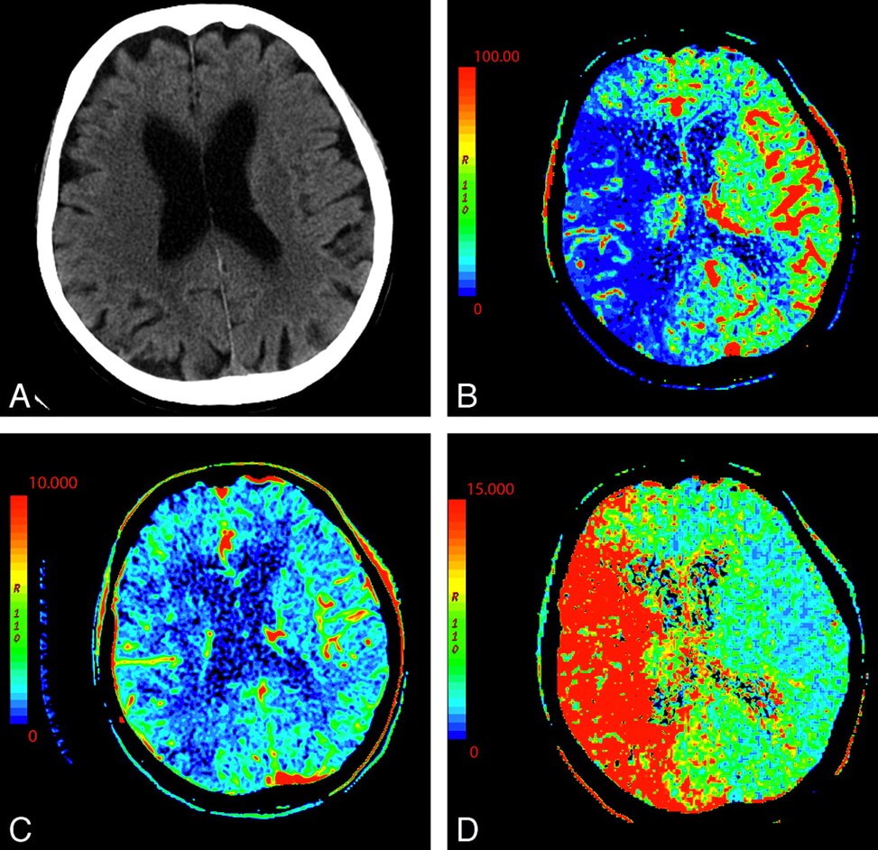
Even with fully automated CT perfusion softwares, clinicians need to learn how to use it rapidly. CT perfusion is determined from imaging of the time-density curves during wash-in and wash-out periods of iodinated contrast injected IV. Multiple perfusion maps are generated. Time to maximum intensity of contrast density in each voxel is one; mean transit time, usually around 5 seconds, is another. Cerebral blood flow is measured in ml/100g/min. Normal is 80 for gray matter and 20 for white matter. Cerebral blod volume is expressed in ml/100g, the area with blood vessels.
Achala Vagal et al. Neurology 2019, 93:888-898
The central volume principle says that cerebral blood flow = cerebral blood volume divided by mean transit time. Measurement of 2 will give the third value. There are 2 further concepts that people need to grasp. First the area under the curve of the time-density curves, which reflects arterial flow and tissue characteristics. Delay or dispersion of contrast bolus may result from injection details, cardiac output, carotid stenosis. In order to avoid these confounding factor, a “deconvolution” mathematical operation allows improved determination of the central volume principle and its key factors.
Relative Cerebral blood flow of 30-45% defines the ischemic core, with delay time to time to maximum intensity, TMax, of >3-6s defines ischemic core, the area destined for infarction. These numbers vary with the software being used. The values indicating delayed endovascular therapy depend on the software and on clinical decisions, reasons why neurolgists need to grasp and operate these concepts rapidly, in order to aquire experience with their teams.
There are significant sources of error. Patient motion, manner of contrast injection, excess radiation are the most important technically, but neurologists need to develop their own evaluation of the software and its calculation of ischemic core. This may be checked with careful consideration of the hypodensity region on standard noncontrast CT imaging. The clinical aim is to minimize under or overestimation of the ischemic core. Miscalculation of the penumbra region is more frequent in the skull base, posterior fossa and orbits, obviously.
Stroke mimics, which appear to be stroke ischemia but are not are seizures, hypertensive encephalopathy and hemiplegic migraine. Especially postictal seizure state is quite similar to the hypoperfusion of stroke. CT perfusion is well defined for the anterior circulation. In the posterior circulation there are artifacts, and diagnostic values have not been defined. Posterior circulation, lacunar and watershed infarcts are much better seen on MRI-DWI than on CT. But MRI has its own difficulties.
Dr Paulo Bittencourt